作者:Zhang, FX; Ye, Q; Chen, QL; Yang, K; Zhang, DY; Chen, ZR; Lu, SS; Shao, XP; Fan, YX; Yao, LM; Ke, LN; Zheng, TL; Xu, H
影响因子:3.62
刊物名称:APPLIED AND ENVIRONMENTAL MICROBIOLOGY
出版年份:2018
卷:84(19)
Prorocentrum donghaiense blooms occur frequently in the Yangtze River estuary and the adjacent East China Sea. These blooms have damaged marine ecosystems and caused enormous economic losses over the past 2 decades. Thus, highly efficient, low-cost, ecofriendly approaches must be developed to control P. donghaiense blooms. In this study, a bacterial strain (strain Y42) was identified as Paracoccus sp. and was used to lyse P. donghaiense. The supernatant of the strain Y42 culture was able to lyse P. donghaiense, and the algicidal activity of this Y42 supernatant was stable with different temperatures and durations of light exposure and over a wide pH range. In addition to P. donghaiense, Y42 showed high algicidal activity against Alexandrium minutum, Scrippsiella trochoidea, and Skeletonema costatum, suggesting that it targets primarily Pyrrophyta. To clarify the algicidal effects of Y42, we assessed algal lysis and determined the chlorophyll a contents, photosynthetic activity, and malondialdehyde contents of P. donghaiense after exposure to the Y42 supernatant. Scanning electron microscopy and transmission electron microscopy analyses showed that the Y42 supernatant disrupted membrane integrity and caused algal cell breakage at the megacytic zone. Photosynthetic pigment loss and significant declines in both photosynthetic efficiency and the electron transport rate indicated that the Y42 supernatant damaged the photosynthetic system of P. donghaiense. Malondialdehyde overproduction indicated that the Y42 supernatant caused lipid peroxidation and oxidative damage to membrane systems in the algal cell, ultimately leading to death. The findings of this study reveal the potential of Y42 to remove algal cells from P. donghaiense blooms.
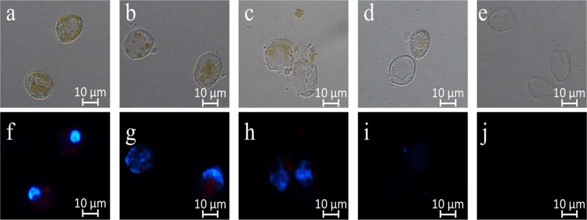
Figure 4. Changes in the morphology and nucleus of P. donghaiense cells during cell death. Algal cells were harvested by centrifugation after 0 h (a and f), 12 h (b and g), 24 h (c and h), 48 h (d and i), or 72 h (e and j) of exposure to 5% of the Y42 strain supernatant, stained with DAPI (5 g/ml) for 5 min at room temperature in the dark, and viewed with a fluorescence microscope. Morphological changes were visualized using bright-field illumination (a to e), and nucleus changes were visualized using UV illumination (f to j).