He CY, CG Wang, BW Li, MF Wu, H Geng, YX Chen and ZH Zuo.Aquatic Toxicology, 2012. 116: 109-115.
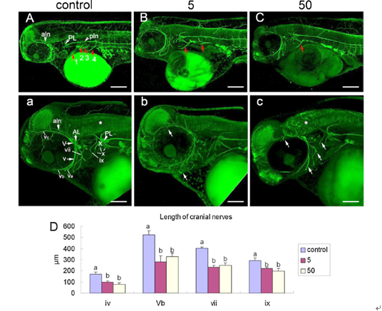
Polycyclic aromatic hydrocarbons (PAHs) are widespread environmental contaminants, which are known to be carcinogenic and teratogenic. These compounds cause a range of macroscopic malformations, particularly to the craniofacial apparatus and cardiovascular system during vertebrate development. However, little is known concerning microscopic effects, especially on the sensitive early life stages or on the molecular basis of developmental neurotoxicity. Using the rockfish (Sebastiscus marmoratus), we explored the neurodevelopmental defects caused by early-life exposure to environmentally relevant concentrations of pyrene, a 4-ring PAH. The results showed that pyrene substantially disrupted the cranial innervations pattern and caused deficiency of motor nerves. The expression of a protein associated with axon growth, growth associated protein 43, was decreased in the central nervous system after treatment with pyrene. N-methyl-d-aspartate receptor (NMDAR) plays a vital role in a variety of processes, including neuronal development, synaptic plasticity, and neuronal survival and death. Our results showed that the expression of Ca2+/calmodulin dependent kinase II and cAMP-response element-binding, which belong to the NMDAR pathway, were increased in a dose-dependent manner after exposure to pyrene. Acetylcholine, an important neurotransmitter which is known to suppress retinal cells neurite outgrowth, was increased by pyrene exposure. Nitric oxide (NO) acts as an activity-dependent retrograde signal that can coordinate axonal targeting and synaptogenesis during development. The level of NO was decreased in a dose-dependent manner following exposure to pyrene. Taken together, the defects in neurodevelopment and the damage to related mechanisms provided the basis for a better understanding of the neurotoxic effects of pyrene.

Fig. 1 Pyrene caused nervous system development defects ofSebastiscus marmoratusembryos as observed by immunostaining of acetylated α-tubulin. (A-C) are 10-fold magnifications of head and trunk and (a-c) are 20-fold magnifications of head. (D) The length of some cranial nerves are measured and analyzed. Data are presented as means ±S.E. (n = 8). Treatments not sharing a common letter are significantly different atP< 0.05 as assessed by one-way anova followed by the tukey hsd test.Abbreviations: aln, anterior lateral line nerve; AL, anterior lateral line ganglion; pln, posterior lateral line nerve; PL, posterior lateral line ganglion; V, trigeminal ganglion; v, trigeminal nerve; Va, upper mandible branch of the trigeminal nerve; Vb, upper jaw branch of the trigeminal nerve; Vc, anterior projecting branch of the trigeminal nerve; vii, facial nerve; ix, hypoglossal nerve; X, vagal ganglion; x, branches of the vagal nerve. Lateral views; anterior to the left. Scale bar A-C = 200µm; a-c = 100 µm

