Sun Y., Zhang Q.J., Zhong J., Wang Y.Q. * Development Growth and Differentiation, 2010.52:157-167.
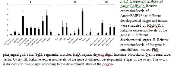
Bonemorphogenetic proteins (BMPs) are responsible for regulating embryo development and tissue homeostasis beyond osteogenesis. However, the precise biological roles ofBMP3andBMP3bremain obscure to a certain extent. In this study, we cloned an orthologous gene (AmphiBMP3/3b) from amphioxus (Branchiostoma japonicum) and found its exon/intron organization is highly conserved. Further,in situhybridization revealed that the gene was strongly expressed in the dorsal neural plate of the embryos. The gene also appeared in Hatschek's left diverticulum, neural tube, preoral ciliated pit and gill slit of larvae, and adult tissues including ovary, neural tube and notochordal sheath. Additionally, real-time quantitative PCR (RTqPCR) analysis revealedthat the expression displayed two peaks at gastrula and juvenile stages. These results indicatedthatAmphiBMP3/3b, a sole ortholog of vertebrateBMP3andBMP3b, might antagonize ventralizingBMP2orthologous signaling in embryonic development, play a role in the evolutionary precursors of adenohypophysis, as well as act in female ovary physiology in adult.


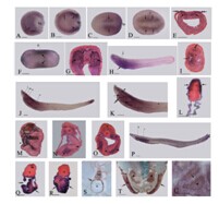
Fig. 4. Developmental expression of AmphiBMP3/3b in the embryo, early larva and adult. In the whole mounts, anterior is on the left; the cross sections are counterstained with pink and viewed from the posterior end of the animal. Scale bars = 50 μm in A–D, F, H, J, K and P; 25μm in E, G, I, L-O, Q and R; 250 μm in S, T and 100 μm in U. (A) Side view of the whole mount of midgastrula (4 hours post-fertilization) with AmphiBMP3/3b expression in the invaginated hypoblast (arrow). (B) Side view of the whole mount of late gastrula (6 hours post-fertilization) with expression throughout the endoderm (double arrow) and in the blastoporal lip (arrows) conspicuously. (C, D) Dorsal view of whole mount of early neurula (7 hours post-fertilization). The expression level is strongest at the neural plate (arrows). (E) Cross section through level e in (D). Transcript is visible at the neural plate (double arrows) and the dorsal mesoderm (arrows). (F) Dorsal view of middle neurula (10 hours post-fertilization). Arrows indicate the expression in the somites. (G) Cross sections through levels g in (F). Arrows indicate the expression in the somites and arrowheads indicate the expression in the neural plate. (H) Side view of early larva in which the mouth has just opened (35 hours post-fertilization). AmphiBMP3/3b is expressed in the pharynx (arrow). (I) Cross section through level i in (H). Arrow indicates the Hatschek's left diverticulum and double arrow indicates the neural tube. (J) Side view of 3-gill-slit larva (6 days post-fertilization) showing the expression in the anterior ectoderm, cerebral vesicle, preoral ciliated pit, club-shaped gland, gill slit, lateral and ventral mesoderm, anterior neural tube and posterior end of the tail. (K) Anterior enlargement of preceding specimen showing transcripts in the cerebral vesicle (arrowhead), preoral ciliated pit (arrow), club-shaped gland (double arrow) and gill slit (asterisk). (L-O) Cross sections through levels l, m, n and o in (J). Arrows indicate the anterior ectoderm in (L), cerebral vesicle in (M), club-shaped gland in (N) and lateral mesoderm in (O). Arrowheads indicate the preoral ciliated pit in (M), endostyle in (N) and neural tube in (O). Double arrows indicate the neural tube in (N) and ventral mesoderm in (O). Asterisk indicates incipient pharyngeal slit in (O). (P) Side view of late larva (33 days post-fertilization) showing the expression in the mouth and ectoderm surrounding the gill slits. (Q, R) Cross sections through levels q and r in (P). Arrows indicate the ectoderm in (Q) and (R). Double arrow indicates the mouth in (Q). (S, T) Expression patterns of AmphiBMP3/3b in adult tissues. Transcripts are visible in the nerve cord, sheath surrounding the notochord (S), metapleural fold and ovary (T). Arrows indicate the sheath surrounding the notochord in (S) and the metapleural fold in (T). Asterisk indicates the ovary in (T). (U) Cross section of ovary. The gene is expressed in oocytes (asterisk). Arrow indicates follicular cells. Abbreviations: bp, blastopore; n, notochord; nc, neural cord; ps, pigment spot.


