Zhong Y.-H.; Guo Z.-J.; Wei M.-Y.; Wang J.-C.; Song S.-W.; Chi B.-J.; Zhang Y.-C.; Liu J.-W.; Li J.; Zhu X.-Y.; Tang H.-C.; Song L.-Y.; Xu C.-Q. and Zheng H.-L. 2023. Plant Cell and Environment 46(5): 1521-39.
Hydrogen sulfide (H2S) is considered to mediate plant growth and development. However, whether H2S regulates the adaptation of mangrove plant to intertidal flooding habitats is not well understood. In this study, sodium hydrosulfide (NaHS) was used as an H2S donor to investigate the effect of H2S on the responses of mangrove plant Avicennia marina to waterlogging. The results showed that 24-h waterlogging increased reactive oxygen species (ROS) and cell death in roots. Excessive mitochondrial ROS accumulation is highly oxidative and leads to mitochondrial structural and functional damage. However, the application of NaHS counteracted the oxidative damage caused by waterlogging. The mitochondrial ROS production was reduced by H2S through increasing the expressions of the alternative oxidase genes and increasing the proportion of alternative respiratory pathway in the total mitochondrial respiration. Secondly, H2S enhanced the capacity of the antioxidant system. Meanwhile, H2S induced Ca2+ influx and activated the expression of intracellular Ca2+ -sensing-related genes. In addition, the alleviating effect of H2S on waterlogging can be reversed by Ca2+ chelator and Ca2+ channel blockers. In conclusion, this study provides the first evidence to explain the role of H2S in waterlogging adaptation in mangrove plants from the mitochondrial aspect.

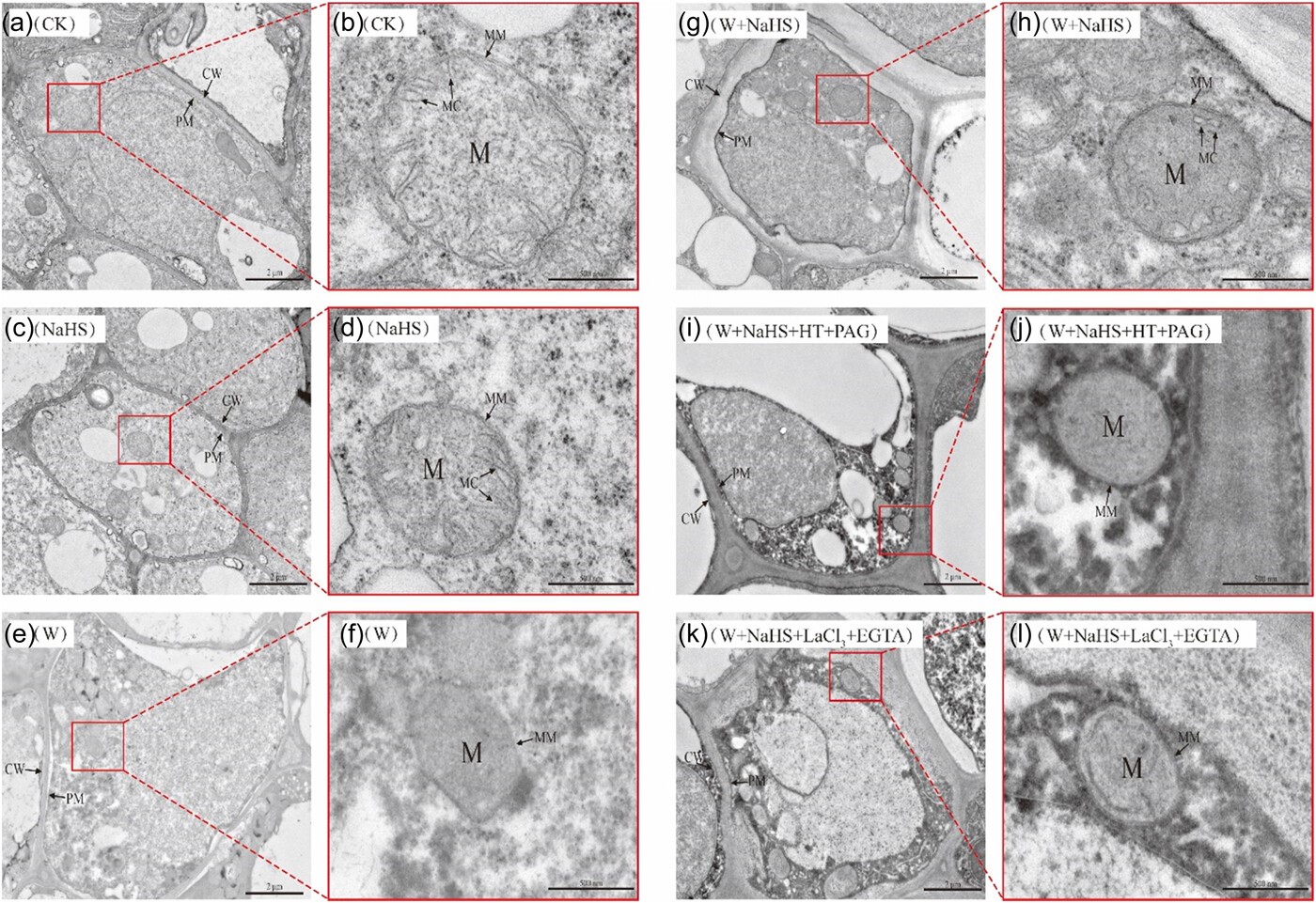
Transmission electron microscope observation of A. marina seedling roots treated with different NaHS under 24-h waterlogging. (a, c, e, g, i, k) The ultrastructure of mitochondria in the root tip cells under low magnifications. Bars in a, c, e, g, i and k represent 2 μm. (b, d, f, h, j, l) The ultrastructure of mitochondria in the root tip cells under high magnifications. Bars in b, d, f, h, j and l represent 0.5 μm. CW, cell wall; PM, plasma membrane; M, mitochondrion; MC, mitochondrial cristae. Other abbreviations are as same as in Figure 3. [Color figure can be viewed at wileyonlinelibrary.com]

